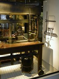
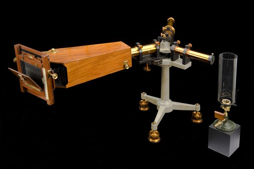
Hilger wavelength spectroscope, with camera, London, England, 1919
- maker:
- Adam Hilger and
- Adam Hilger Limited
![]() This image is released under a CC BY-NC-SA 4.0 Licence
This image is released under a CC BY-NC-SA 4.0 Licence
Buy this image as a print
BuyLicense this image for commercial use at Science and Society Picture Library
License1981-151, Hilger wavelength spectrometer, with camera, by A
Science Museum Group Collection
© The Board of Trustees of the Science Museum
Hilger wavelength spectrometer, with camera, by A. Hilger, London, 1918-1920
This spectroscope is used to study which light waves from the spectrum are absorbed when passed through living body tissue. The camera records the wavelengths that have been absorbed by taking a spectrograph.
This spectroscope was presented to Royal Wolverhampton hospital in recog-nition for the work of their first honorary pathologist, Charles Alexander MacMunn (1852-1911). MacMunn used a spectroscope to demonstrate the existence of a particular pigment in muscle, which plays an important role in cellular respiration similar to that of haemoglobin. These are known as cytochromes. He also wrote 'The Spectroscope in Medicine' in 1880. The spectroscope is shown here with a Bunsen burner (1905-103/2).
Details
- Category:
- Laboratory Medicine
- Object Number:
- 1981-151
- Materials:
- camera, mahogany, spectrometer, brass and spectrometer, steel
- Measurements:
-
overall: 450 mm x 830 mm x 515 mm,
- type:
- spectroscope
- credit:
- New Cross Hospital