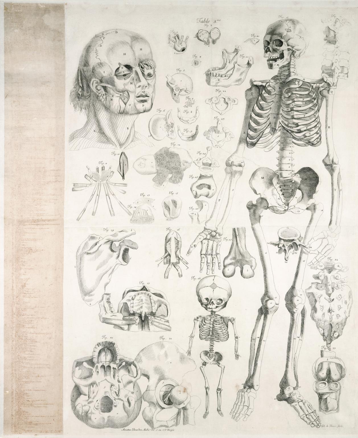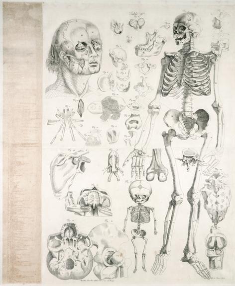
Print showing the musculature of the human body, Paris, France, 1678
print; engraving, with Mss. keys added lhs. Anatomical drawing /Ame Bourdon; engraved by Daniel le Bossu; published by Laurens D'Houry, Paris 1678. 110x91.5cm. Being Table 1 (of 16) from A Bourdon, Nouvelle description Anatomique (Paris 1678)
Showing the male human body complete with the muscles and skin, this anatomical drawing was one of sixteen plates of 'Nouvelles tables anatomiques' or 'New Anatomical Tables'. Each plate was drawn by Amé Bourdon (1683-1706), a French physician and anatomist, and engraved by Daniel Le Bossu. On the left hand side is a manuscript key to identify each part of the body. The work was published in 1679. The close relationship between art and anatomy began in the Renaissance, when artists and anatomists worked closely together to produce detailed anatomical drawings for medical textbooks, and established artistic and aesthetic conventions.