![]() This image is released under a CC BY-NC-SA 4.0 Licence
This image is released under a CC BY-NC-SA 4.0 Licence
Buy this image as a print
BuyLicense this image for commercial use at Science and Society Picture Library
LicenseWooden anatomical figure, female, possibly 17th century
Science Museum Group Collection
© The Board of Trustees of the Science Museum
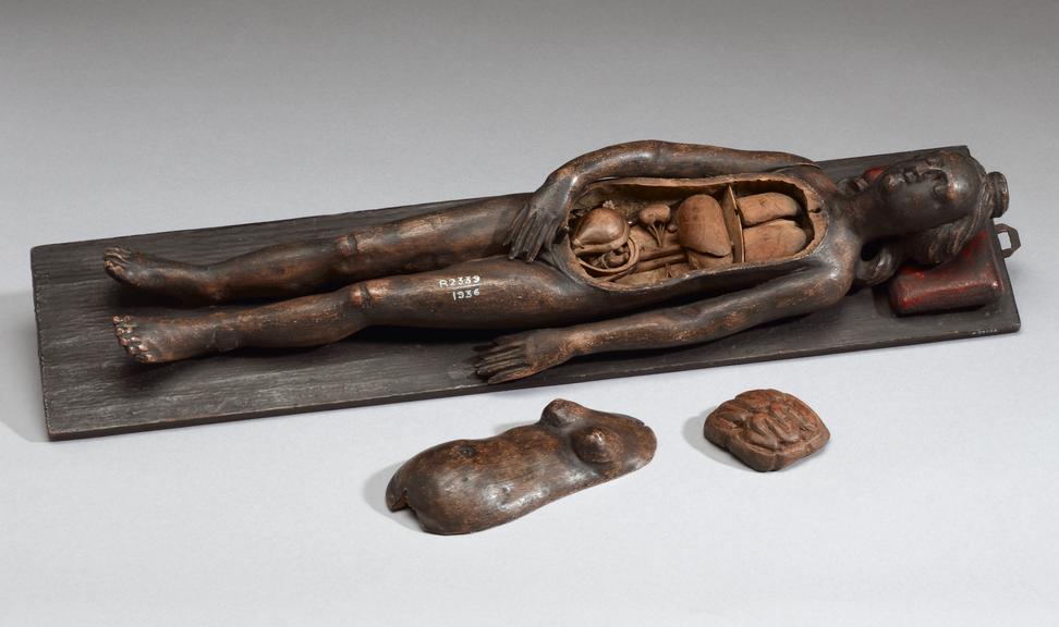
Wooden anatomical figure, female, possibly 17th century
The torso of this figure can be removed to show the intestines, liver, stomach and a basic representation of the lungs and diaphragm. The intestines are also removable to show the reproductive organs. The uterus can be seen and when the flap is removed a foetus is revealed. The underside of the removable torso is engraved with muscles and arteries.
Many anatomical figures from this period tended to be made of ivory and were considerably smaller. This model is 430 mm in length. The level of anatomical detail is limited but gives a basic layout of the main organs. As such it is likely that the model was used to teach lay people about basic human anatomy. It may possibly have been used by midwives to provide reassurance for pregnant women and to teach young married couples about anatomy and pregnancy. This model forms a pair with A79252 – a male figure. Pairs of male and female figures were not uncommon in the 1600s.
Details
- Category:
- Anatomy & Pathology
- Collection:
- Sir Henry Wellcome's Museum Collection
- Object Number:
- A79253
- Materials:
- wood
- Measurements:
-
overall: 80 mm x 100 mm x 433 mm, .5kg
- type:
- anatomical figure
- credit:
- Bernard, H.