![]() This image is released under a CC BY-NC-SA 4.0 Licence
This image is released under a CC BY-NC-SA 4.0 Licence
Buy this image as a print
BuyLicense this image for commercial use at Science and Society Picture Library
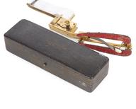
LicenseBronchoscope, rigid, Fourestier-Glandu-Vulmiere type
Science Museum Group Collection
© The Board of Trustees of the Science Museum
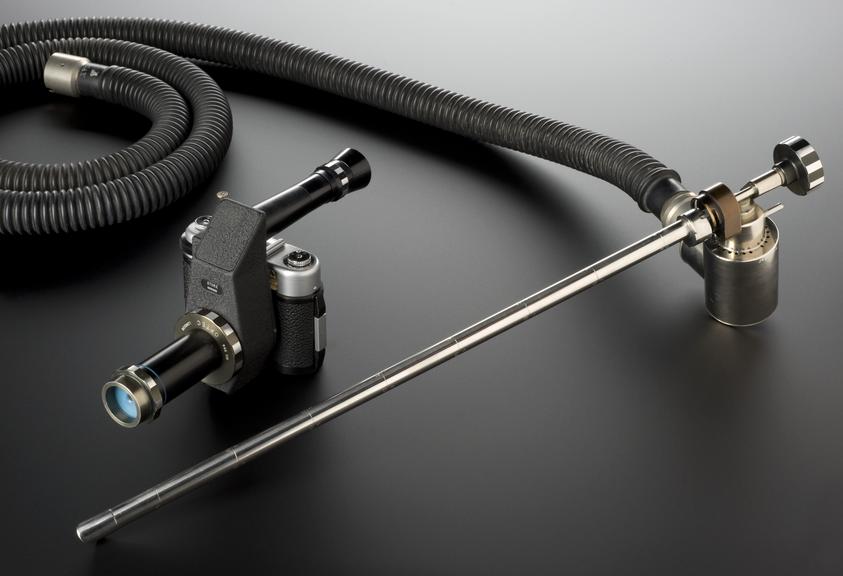
Hysteroscope, rigid, Fourestier-Glandu-Vulmiere type, with camera attachment, France, 1958-1965
Physicians ‘saw’ into the body using a hysteroscope. A rigid tube is passed into the body to 'see' inside the uterus. This example incorporated a quartz rod within the tube. Light was transmitted by an internal reflection in the rod. This meant the lens systems of earlier bronchoscopes were discarded. Illumination was good enough for a specially mounted camera to take very clear photographs of the interior of the uterus.
Details
- Category:
- Clinical Diagnosis
- Object Number:
- 1981-952
- Materials:
- steel (stainless), rubber, metal, brass (nickel plated), paper (fibre product), plastic (unidentified) and glass
- Measurements:
-
overall: 130 mm x 115 mm x 240 mm, 1.46 kg
overall (as displayed): 130 mm x 600 mm x 310 mm, 2.382 kg
- type:
- hysteroscope


