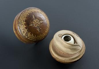
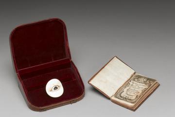
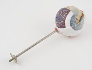
Anatomical model of the eye with a descriptive booklet in a leather case, Italian, 1674-1679 1674-1679
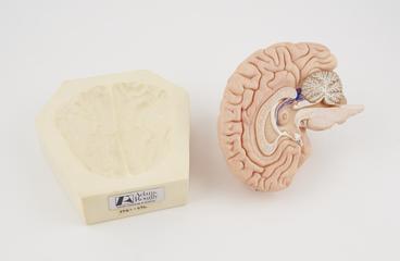
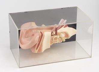
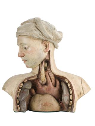
Wax anatomical dissection model of human head showing internal structure of brain, eye, cheek, neck and jaw, unsigned, European (possibly German), 1801-1900 (see note). Wax anatomical model of a human head 1801-1900
Anatomical model of human foot bones, origins unknown, part of a chirpodist's collection Anatomical model of human foot bones before 1988
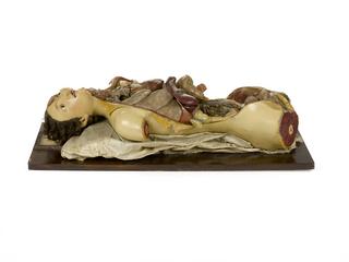
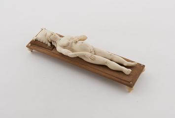
18th century wax anatomical preparation, probably by Fragonard 18th century wax anatomical preparation 1701-1800
Wax model of human face showing internal structure of eye, German(?), 19th century Wax model of a human face, Germany, 1801-1900 1801-1900
Two life size wax anatomical heads, illustrating the effects of secondary syphilis and their treatment with Salvarsan, preparation 606, mounted in a glass display case, probably made in Germany, 1910-1920 Two wax heads showing the effects of syphilis and their treatment, Germany, 1910-1920 1910-1920
Full-size female animated figure model, part of installation with motorised base and four hear phones through which recording of narration about the human body can be played, from Cleveland Health Museum, Ohio, United States, 1965 Female animated figure model from Cleveland Health Museum, Ohio, United States, 1965 1965
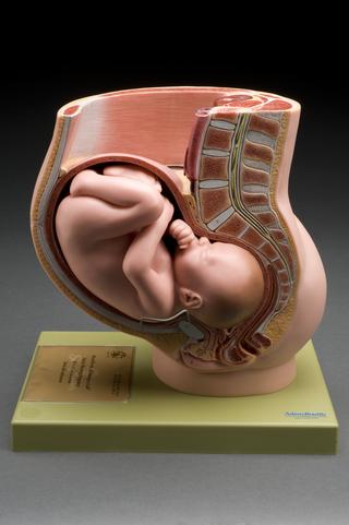
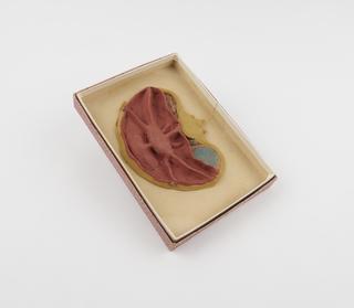
Full scale cut away anatomical model of a female abdomen showing 12-week foetus in the womb, by Dr. P. Freeborn Ltd., British Cut away anatomical figure of a female abdomen, 1977. 1977
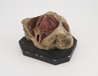
Anatomical model of the human heart on stand with section to show main valves from above and below (12" tall) anatomical model; heart
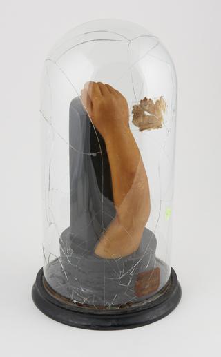
Three life size wax leg segments showing stages in the successful treatment of ulcers caused by secondary stage syphilis with Salvarsan, preparation 606, mounted glass display case, with original wooden packing crate, probably made in Germany, 1910-1920 Three wax legs showing treatment of ulcers caused by syphilis, Germany, 1910-1920 1910-1920
Two life size wax anatomical heads, illustrating the effects of tertiary syphilis and their treatment with Salvarsan, preparation 606, mounted in a glass display case, probably made in Germany, 1910-1920 Two wax heads showing the effects of syphilis and their treatment, Germany, 1910-1920 1910-1920
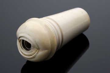
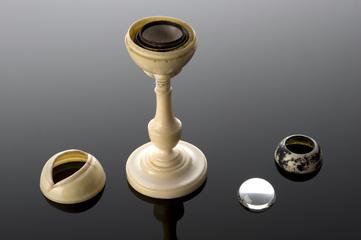
The Ho model eye demonstrating basic principles of vision. With a brass base and pillar supporting tube fitted with the iris diaphragm. There is a provision for inserting glass lenses, ground on rear surface, and different curvatures on front face. The lenses are marked I, II, III, and IIII. F.E. Becker and Co. c.1913 The 'Ho Model' eye 1909-1919